Virtual Patient Workflow in Anterior Smile Design: Clinical and Laboratory Value of 3DIFY Scanning System
In anterior restoration workflows, dental technicians are the core controllers of the final restoration’s morphology, marginal adaptation, aesthetic outcome and soft tissue compatibility. Clinical tooth preparation and impression taking are performed by dentists.
However, numerous remakes of anterior restorations are highly associated with secondary errors occurring in dental laboratories, including model deformation, damage during trimming, and subjective interpretation of the finish line. The long-term prognosis of restorations depends not only on clinical operation quality, but also on the accuracy of data acquisition and morphological reproduction at the laboratory stage.
Currently, digital anterior restoration in the market mostly relies on intraoral scanning and desktop model scanning to obtain hard tissue data of the dentition, which enables good control of marginal adaptation. Nevertheless, relying solely on dental hard tissue data has limitations in matching restorations with facial aesthetics. The size, inclination and incisal morphology of teeth need to be harmonized with the patient’s facial form, lip line, smile line and facial symmetry. Pure dentition scan data lacks facial references, which may lead to aesthetic failures where the teeth look well-formed yet disharmonious when placed on the patient’s face.
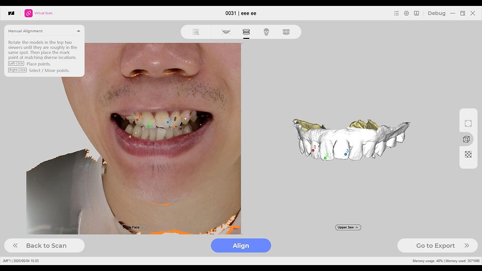
Facial scanning fills this gap: it captures the patient’s 3D facial features, smile arc, lip-tooth relationship and facial midline. The facial soft tissue data can be registered and fused with the dental and jaw hard tissue model acquired via desktop model scanning, realizing integrated facial-dental digital design.
Desktop model scanners deliver high scanning precision to eliminate human errors caused by manual observation of traditional plaster models. They stably reproduce finish line contours, interdental spaces and occlusal morphology, greatly reducing common laboratory issues such as poor marginal adaptation, over-tight or over-loose proximal contacts and occlusal high spots, and improving the fundamental precision of restorations. Facial scanning provides facial reference data, allowing technicians to design anterior tooth morphology, shade, alignment and incisal curve based on the patient’s real facial features rather than only observing dental models. This balances functional adaptation and smile aesthetics.
1. Core Technical Points of Anterior Aesthetic Restoration
1.1 Reproduction of Finish Line and Control of Marginal Adaptation
The long-term prognosis of anterior restorations fundamentally relies on marginal adaptation. Traditional plaster models are prone to deformation during pouring, transportation and trimming. Manual observation of finish line boundaries carries strong subjectivity, and tiny defects can easily be overlooked.
Desktop model scanning rapidly generates high-precision 3D models, clearly identifying continuous finish line contours and digitally marking marginal lines to reduce deviations from manual model trimming, laying a foundation for well-adapted crowns, veneers and other anterior restorations.
1.2 Smile Aesthetic Design Requires Reference to Facial Soft Tissues
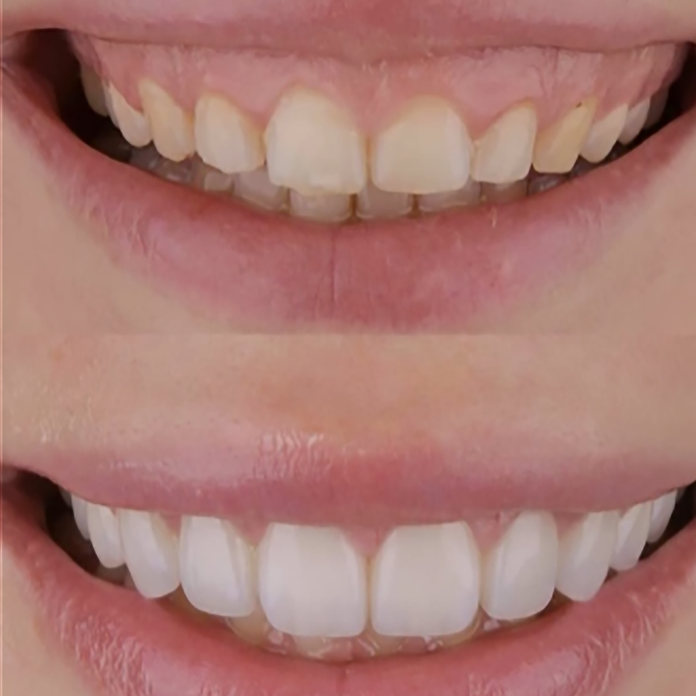
Traditional digital workflows only provide dentoalveolar data. Technicians can only design based on 2D photographs, which suffer from perspective distortion and angle deviation. It is difficult to precisely align the midline, smile line and lip-tooth gap.
3D facial scanning outputs a three-dimensional facial model that can be imported directly into restoration design software:
Extract facial midline, pupillary line, lip line and smile arc
Evaluate lip-tooth relationship at rest and tooth exposure during dynamic smiling
Match the aspect ratio of anterior teeth and dental arch curve with facial morphology
After data fusion, restoration design is upgraded from beautifying individual teeth to personalized smile rehabilitation, lowering patients’ subjective dissatisfaction caused by disharmonious aesthetics after treatment.
1.3 Prediction and Control of Interdental Spaces and Black Triangles
Black triangles are a common aesthetic concern in anterior restorations, closely related to tooth morphology, the position of proximal contact areas and alveolar soft tissue morphology.
Desktop scanning accurately reproduces proximal tooth morphology. Combined with lip soft tissue contours from facial scanning, technicians can predict the visual effect of interdental spaces and lip compression on teeth in the virtual design stage. The height of contact areas can be properly adjusted to reduce the visual appearance of postoperative black triangles.
1.4 Coordination of Occlusion in Static and Dynamic Functions
Desktop scanning acquires complete occlusal information of the dentition and reconstructs occlusal contact points. Combined with facial scan data, it is possible to assess the movement trajectory of anterior incisal edges during smiling and speaking with consideration of mandibular movement and facial expressions. This prevents restorations from interfering with lip movement and balances function and aesthetics.
2. Improved Laboratory Value of Combined Desktop Model Scanning and Facial Scanning
Precision: Desktop scanning reproduces subtle anatomical structures of plaster models and eliminates errors from manual measurement and trimming, ensuring marginal adaptation of restorations and reducing secondary caries and gingival inflammation caused by marginal gaps.
Communication: The fused 3D facial and dentition model can intuitively display the predicted smile effect after design. It facilitates communication between technicians and clinicians, and helps dentists preview outcomes with patients before treatment, reducing expectation gaps and aesthetic-related remakes.
Prognosis: Improved marginal adaptation reduces periodontal irritation. Tooth alignment and incisal morphology matching the patient’s facial physiological features make restorations more compatible with natural facial expressions, delivering better long-term wearing stability and lowering long-term complications such as gingival redness and ceramic chipping.
3. Overview of Digital Workflow (3DIFY Solution)
Clinical side: Capture 3D facial scan data of the patient and record smile status via facial scanning.
Take impressions and fabricate plaster models, then send models to the dental laboratory.
Laboratory side: Scan plaster models with a desktop model scanner to obtain high-precision 3D dentoalveolar data.
Register and align the facial model and dentition model within design software.
Carry out virtual design for anterior veneers, full crowns and other aesthetic restorations based on fused data, simultaneously considering margin, occlusion and smile aesthetics.
Export data for milling or 3D printing to fabricate restorations.
For anterior aesthetic restoration, functional marginal adaptation is the foundation, while facial harmony constitutes the core of aesthetics. Desktop dental model scanning alone can only solve hard tissue precision challenges and hardly predict the real smile effect of restorations on patients’ faces.
The 3DIFY solution combining desktop model scanning and facial scanning connects the digital workflow of facial soft tissues and dentoalveolar hard tissues. Dental technicians can not only acquire high-precision model data to accurately reproduce finish lines, interdental spaces and occlusion, but also conduct personalized smile design relying on 3D facial information. It reduces remakes caused by poor marginal adaptation and aesthetic disharmony from the source, and enhances aesthetic performance and long-term clinical prognosis of anterior restorations.